Blog

Dehydroepiandrosterone sulfate
22 October 2025
Dehydroepiandrosterone sulfate (DHEA-S) is a steroid hormone synthesized in the adrenal cortex’s reticular zone under the influence of adrenocorticotropic hormone (ACTH). Like other steroids, DHEA-S is synthesized from cholesterol. DHEA-S is considered a prohormone. It is hormonally inert but can be converted into other, more potent androgens and estrogens. It is an important marker of adrenal function and is useful for diagnosing various endocrine disorders, as well as for evaluating premature adrenarche in children. During fetal development, DHEA-S is produced in the adrenal glands; its level rapidly decreases during the first year of life and then rises again at adrenarche (around 6–8 years old), gradually increasing during puberty and reaching its peak between the ages of 20–30. After that, DHEA-S levels steadily decline. In males, the adrenal glands contribute only a small portion of total androgen production, while in women of reproductive age, the adrenal glands play a more significant role in androgen production. In women, elevated DHEA-S levels may cause signs of hyperandrogenism (hirsutism, acne, virilization). Men are usually asymptomatic, but due to peripheral conversion of androgens to estrogens, a mild excess of estrogens may sometimes be observed. Common causes of elevated DHEA-S levels include: 1. Androgen-secreting adrenal tumors (adenomas or carcinomas), especially when DHEA-S is markedly elevated (more than 5 times above normal). 2. Congenital adrenal hyperplasia. 3. Polycystic ovary syndrome (PCOS), often with mild to moderate elevation. Common causes of low DHEA-S levels include: 1. Adrenal insufficiency, such as Addison’s disease or secondary adrenal insufficiency due to hypopituitarism. 2. Subclinical or overt hypercortisolism. 3. Severe systemic illnesses, acute stress, or chronic conditions such as anorexia. DHEA-S affects immune modulation, inflammation, and neuronal protection; low levels are associated with cognitive decline and mood disorders. It is important to regularly monitor the hormone level, as its deviations may indicate adrenal or hormonal disorders at an early stage. Timely DHEA-S testing contributes to accurate diagnosis and the selection of effective treatment.
Respiratory viral infections in children
20 October 2025
With the beautiful autumn days and colors, upper respiratory tract infections or acute respiratory viral infections (ARVI) become more frequent. People often call them seasonal colds. Let’s understand together how children get infected with these viruses and what should be done to prevent them. Upper respiratory tract infections are seasonal viral diseases transmitted by airborne droplets (coughing/sneezing on each other, touching household items/toys of an infected person, using the same dishes). These infections are self-limiting and usually start in autumn, continuing until late spring (or, as they say, along with the school year). The main symptoms are: • Sudden and frequent fever (38°C and above) • Chills, shivering • Headache, muscle pain, body aches • Sore throat • Cough • Runny nose • Sometimes vomiting or diarrhea Respiratory viruses mainly affect the upper respiratory tract — the nasopharynx, throat, and trachea — but sometimes can also involve the bronchi. The illness usually lasts up to one week, sometimes a little longer. What to do in case of viral infection When a child gets infected, the main goal is to ease their condition and prevent complications. • Offer plenty of fluids (water). • Clean the nasal passages with saline solution (NaCl 0.9%) or sea salt sprays. • In case of fever, give Acetaminophen (Paracetamol) or Ibuprofen (Nurofen) in syrup, suppository, or tablet form — according to age and weight, and only as prescribed by a pediatrician. • Ensure constant room ventilation and maintain air humidity. • In case of cough, honey can be useful for children over 1 year old (it is not allowed for younger ones due to the risk of botulism). Important facts about medication Ibuprofen should not be used for dehydrated or persistently vomiting children. Aspirin is prohibited under the age of 12. Antiviral suppositories (Viferon, Genferon) have no proven effectiveness and are often confused with fever reducers, which can be dangerous during high fever. The only antiviral medication with proven effect is Oseltamivir (Tamiflu), used in confirmed influenza cases, starting from the first day of illness and only by pediatric prescription. When to seek immediate medical attention If you notice that your child has: • Frequent or labored breathing • Cyanotic (bluish) lips • Retractions between the ribs while breathing • Chest pain • Severe muscle pain causing refusal to walk • Signs of dehydration (no urination for 8 hours or more, dry mouth, crying without tears) • Weakness or lack of response • Convulsions • Persistent high fever (40°C and above) • Age under 3 months • Worsening or recurrence of symptoms after a few days • Presence or exacerbation of chronic diseases (e.g. diabetes, asthma) To prevent respiratory viral infections, it is important to teach children to wash their hands frequently (especially after returning home), eat healthy and balanced meals (with plenty of fruits and vegetables), avoid contact with sick individuals, and receive all vaccinations according to the national immunization schedule (including the seasonal flu vaccine). Thus, follow and teach your children to maintain hygiene rules, eat healthy and varied food, have sufficient physical activity, and good sleep.
What to do if your child is coughing
17 October 2025
Cough is a natural protective reflex of the body and plays an important role in the innate immunity of the respiratory system. It helps clear mucus, microorganisms, and irritants from the airways. The mechanism of coughing includes three phases: inhalation, compression, and exhalation. The effectiveness of a cough depends on several factors: • the proper width of the airways (narrowing reduces its efficiency), • the properties of mucus, • and the strength of the respiratory muscles. It’s very important not to suppress the cough reflex without identifying and treating the underlying cause. Cough occurs in almost all children, including premature infants (about 10% of babies born at 27 weeks and up to 90% of full-term newborns). It is one of the main clinical signs of respiratory diseases, but it can also appear in non-respiratory conditions. Types of Cough • Normal (expected) — during viral infections • Specific — with sudden shortness of breath, recurrent pneumonia, neuromuscular disorders, or chest/jaw anomalies • Nonspecific — dry cough without an identified respiratory disease (may resolve spontaneously) Cough is also classified by: 1. Cause 2. Duration: Acute — up to 4 weeks Chronic — longer than 4 weeks 3. Nature: Dry or wet (productive) Acute Cough In children, acute cough is most often associated with viral infections of the upper respiratory tract. It usually resolves on its own after the infection and rarely requires further testing. If the cough is persistent, lasts more than 4 weeks, or is accompanied by other concerning symptoms, a doctor’s visit is necessary. Acute cough may also develop after foreign body aspiration, especially in children aged 3–5 years, who tend to put small objects in their mouths. This situation requires immediate medical attention. Acute cough can indicate both respiratory and non-respiratory diseases. Respiratory causes include: nasopharyngitis, sinusitis, false croup, tracheitis, bronchitis, pneumonia, and asthma. Non-respiratory causes include neurological problems, congenital heart defects, and others. The most common cause of acute cough is the common cold, accompanied by sneezing, fever, runny or blocked nose, headache, and sore throat. These are usually self-limiting viral infections that don’t require antibiotics. However, if the child breathes rapidly, has a persistent fever, weakness, or refuses fluids — medical evaluation is necessary. Chronic Cough Chronic cough lasts more than 4 weeks and may be accompanied by wheezing, burning, breathing difficulty, nasal congestion, or hoarseness. You should see a doctor if your child has: • frequent, persistent cough, • barking or nighttime cough, • wheezing or noisy breathing, • productive cough with mucus, • rough or hoarse-sounding cough. Chronic cough may occur in both younger and older children. • In children under 5 years old, it can be caused by infections, asthma, foreign body aspiration, passive smoking, or congenital abnormalities. • In children over 5 years old, causes include asthma, prolonged bacterial bronchitis, allergies, long-term use of nasal drops, and passive smoking. What to Do During autumn, when viral infections are more common, it’s important to pay close attention to cough symptoms and seek timely care. If your child has a cough, don’t wait for it to pass on its own. Consult a pediatrician. Our clinic’s experienced doctors will identify the cause, prescribe the right treatment, and, if necessary, perform modern diagnostic tests. Our clinic offers all the necessary advanced diagnostic methods to ensure accurate diagnosis and effective treatment.
Duplex Examination of the Lower Limb Vessels
16 October 2025
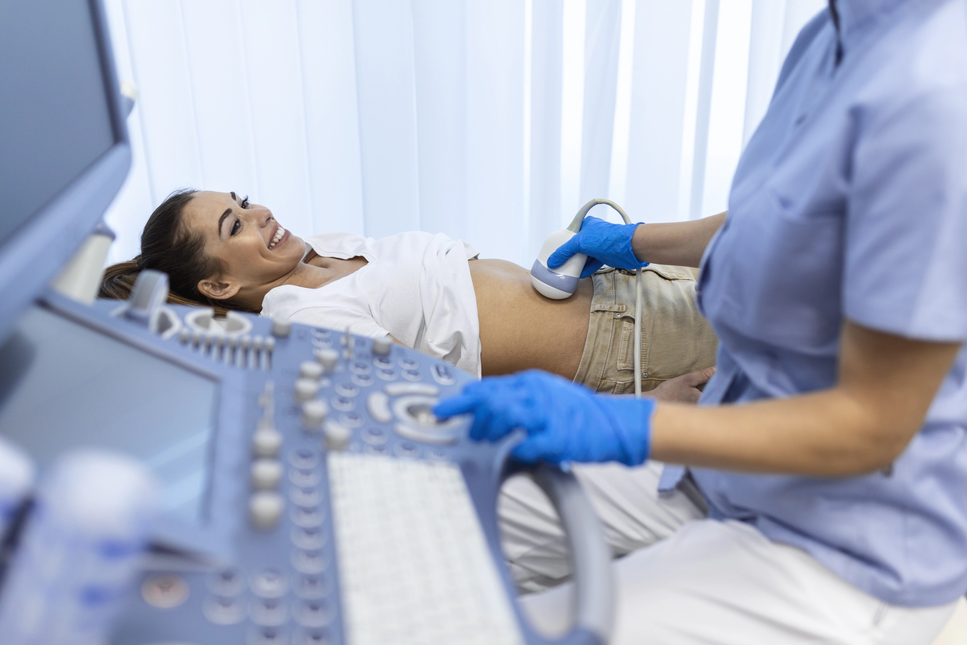
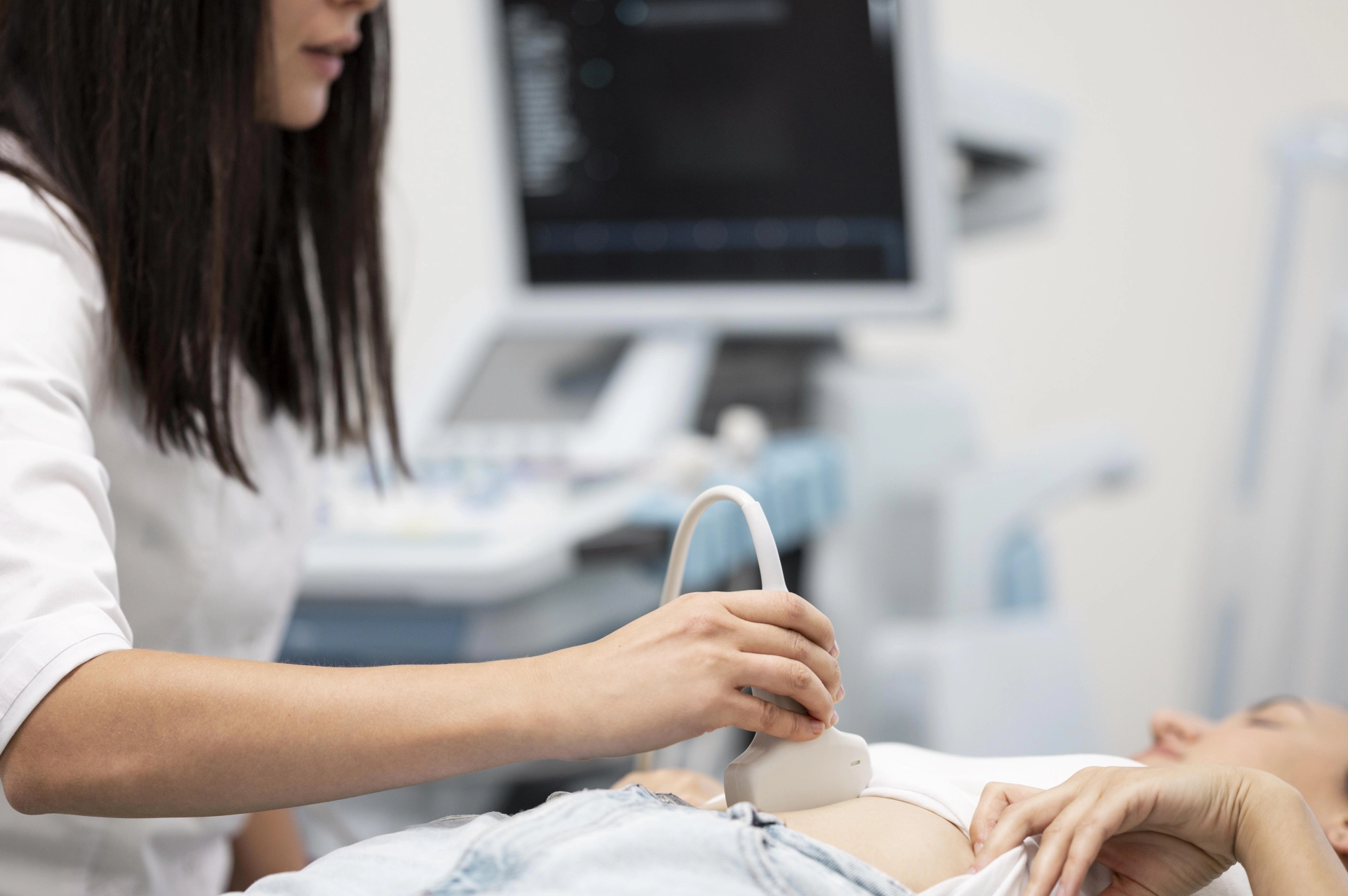
The duplex examination is a modern ultrasound method that allows for the assessment of blood circulation in the lower limbs and the structural condition of the vessels. Through this technique, it is possible to evaluate vessel patency, the degree of narrowing, the state of the vessel wall and lumen, measure vessel diameter, assess blood flow velocity, and detect deformations or the presence of atherosclerotic plaques. Advantages of Duplex Examination • Painless and short (takes about 15–30 minutes) • Absolutely safe (can be performed even during pregnancy) • Affordable and highly informative • No age or gender limitations When the Examination Is Recommended • Swelling of the legs • Feeling of heaviness or discomfort in the lower limbs • Pain or muscle tension during walking (after 10, 50, or 100 meters) • Visible varicose veins • Changes in skin color • Numbness • Vascular “stars” or pronounced subcutaneous vein changes • Diabetes mellitus People at Risk • Those with arterial hypertension • Individuals with cardiovascular diseases • People with obesity • Patients with atherosclerosis or high cholesterol • Individuals with diabetes mellitus • Smokers Duplex examination plays an important role in the early diagnosis of vascular diseases. It allows timely detection of circulation disorders, helps prescribe effective treatment, and prevents the development of complications.
Uterine Cavity Polyp
15 October 2025
An endometrial polyp is a benign growth of the uterine cavity lining. The incidence increases during reproductive age and the perimenopausal period. Main factors contributing to the development of uterine polyps 1. Hormonal imbalance Increased estrogen levels and decreased progesterone levels lead to endometrial hyperplasia (excessive thickening), which contributes to polyp formation. 2. Inflammatory and infectious processes Chronic inflammations, such as endometritis, and infections, including sexually transmitted diseases, can disrupt endometrial cell processes and promote abnormal growth. 3. Mechanical injury Damage to the uterine lining caused by miscarriages or diagnostic curettage may lead to the development of polyps. 4. Endocrine diseases Women suffering from type 2 diabetes, polycystic ovary syndrome (PCOS), or thyroid dysfunction are at higher risk of developing polyps. 5. Excess weight and sedentary lifestyle Obesity and low physical activity affect hormonal balance, which in turn increases the risk of polyp formation. 6. Genetic predisposition Hereditary susceptibility to gynecological diseases and reproductive system tumors may also be a risk factor. 7. Early menarche or late menopause Menstrual onset before age 12 or menopause after the typical age is associated with elevated estrogen levels, which raises the risk of polyp development. Symptoms In many cases, polyps may be asymptomatic, but sometimes the following symptoms occur: • Irregular or intermenstrual bleeding • Heavy menstrual bleeding • Lower abdominal pain • Infertility Diagnosis A polyp can be detected by: • Ultrasound examination (sonography) • Hysterosonography • Hysteroscopy (considered the “gold standard”) Treatment Polyp removal is mainly performed by hysteroscopy, which serves both diagnostic and therapeutic purposes. The removed tissue must be sent for histopathological examination to exclude the presence of malignant cells.Prevention and Monitoring • Regular gynecological examinations • Sonography for early detection • Regulation of hormonal balance Uterine polyps are generally benign and treatable. Timely detection and proper management are crucial for maintaining both women’s health and reproductive function.
Eyelid Demodicosis — Causes, Symptoms, and Modern Treatment Methods
13 October 2025
Demodicosis is a common yet often overlooked inflammatory disease of the eyes and eyelids, caused by the excessive multiplication of microscopic mites — Demodex folliculorum and Demodex brevis — which normally live on the human skin. These mites inhabit the hair follicles and meibomian glands, feeding on skin sebum and dead cells. Under normal conditions they do not cause harm, but when their number exceeds the norm, an inflammatory reaction occurs, leading to irritation, itching, and various ocular surface disturbances. The activation of these mites is related to both internal and external factors. The main causes include: • Weakened immune system due to chronic diseases, infections, or prolonged stress. • Increased skin oiliness, which creates a favorable environment for mite growth. • Hormonal imbalance, such as thyroid or ovarian dysfunction. • Age-related changes, as the condition is more common in middle-aged and elderly people. • Poor eyelid hygiene and overuse of oily cosmetics, especially mascara and eye creams. When several of these factors act together, Demodex mites begin to reproduce rapidly, causing inflammation of the eyelids and eye surface. Symptoms Demodicosis develops gradually. Initially, a person may notice mild itching or tingling around the eyelids, but over time the symptoms become more pronounced. Typical symptoms include: • Itching and burning of the eyelids, especially in the morning. • Redness, swelling, and a feeling of heaviness in the eyelids. • Yellowish-white crusts or discharge at the base of the eyelashes. • Tearing, a gritty or foreign body sensation. • Recurrent inflammation along the eyelid margins, sometimes involving the cornea and conjunctiva. If the mites affect the meibomian glands, meibomian gland dysfunction may develop, causing chronic eye dryness, irritation, and light sensitivity. Diagnosis Diagnosis is confirmed by an ophthalmologist through microscopic examination of eyelashes to identify mites and their eggs. Additionally, the condition of the cornea and tear film is evaluated to determine the severity of inflammation and to detect possible complications. Modern Treatment Approaches Treatment of demodicosis is lengthy and requires a complex, combined approach aimed at reducing mite activity, eliminating inflammation, and restoring normal eyelid function. The main treatment directions are: 1. Eyelid and eyelash hygiene — daily cleansing with special lotions or wipes to remove secretions and mite waste; warm compresses to improve meibomian gland function. 2. Topical therapy — ointments and drops containing anti-mite components such as tea tree oil, metronidazole, or ivermectin; anti-inflammatory agents to reduce redness and swelling. 3. Systemic therapy — oral medications in severe cases, prescribed only under medical supervision. 4. Supportive therapy — artificial tears to relieve dryness, as well as lifestyle adjustments including healthy diet, adequate sleep, and stress management. Prevention To prevent recurrence, it is essential to maintain regular eyelid hygiene, limit the use of oily cosmetics, and avoid using old or contaminated makeup products. Any early signs such as itching, redness, or tearing should prompt a visit to an ophthalmologist to avoid complications. Conclusion Demodicosis does not threaten vision directly, but it can significantly affect quality of life by causing chronic discomfort, irritation, and cosmetic concerns. Early diagnosis, proper hygiene, and comprehensive treatment allow effective control of the disease and help prevent relapses.
Helicobacter pylori
12 October 2025
Helicobacter pylori is a spiral-shaped bacterium that lives and multiplies in the human stomach. With its thin flagella, it easily moves through the stomach’s mucous layer and attaches to it, surviving even in the strong acidic environment of the stomach. Discovery and Importance Helicobacter pylori was first discovered in 1982. Since then, it has been proven that the bacterium is associated not only with gastritis and stomach or duodenal ulcers, but also with certain types of stomach cancer. Transmission The infection is mainly transmitted from person to person: • Through oral contact (for example, when sharing spoons, cups, or dishes), • Or through dirty hands. How It Causes Disease Once inside the stomach’s mucosal layer, Helicobacter pylori produces an enzyme called urease, which neutralizes stomach acid around it. This creates a “safe zone” that allows the bacterium to survive. Over time, it damages the stomach lining, leading to inflammation — gastritis, ulcers, and, in some cases, even cancer. Main Symptoms Although some people may carry Helicobacter pylori without symptoms, common signs include: • Pain in the upper part of the stomach • Heartburn and discomfort after meals • Nausea, sometimes vomiting • Loss of appetite and weight loss • Dark or bloody stools (in case of ulcer bleeding) If Left Untreated If not treated, Helicobacter pylori can cause: • Chronic gastritis • Stomach and duodenal ulcers • Ulcer complications such as perforation or bleeding • Increased risk of stomach cancer Treatment Treatment follows a specific antibiotic-based therapy, often combined with acid-suppressing medications. It usually lasts from 15 days to 1 month and must be completed under a doctor’s supervision. Prevention To prevent Helicobacter pylori infection: • Maintain good personal hygiene — wash hands before and after eating, • Avoid sharing utensils, spoons, and cups, • Eat clean, well-prepared food, • Get regular checkups if you have gastritis or ulcer symptoms. Conclusion Helicobacter pylori is a very common bacterium that may remain unnoticed for years, but over time it can cause serious problems. Early diagnosis and proper treatment are key to preventing complications and protecting your stomach health.
Myocardial Infarction
09 October 2025
Myocardial infarction (MI) is ischemic necrosis of the heart muscle caused by an acute disruption of its blood supply. It is one of the most dangerous cardiovascular diseases and remains one of the leading causes of death worldwide. According to statistics, most cases occur in individuals aged 45–65. Men tend to develop the disease at a younger age than women. In recent years, due to preventive measures in developed countries, the incidence of infarction has somewhat decreased, but it remains a major global health problem. Etiology and Risk Factors The main cause of myocardial infarction is atherosclerotic damage to the coronary arteries. After rupture of an atherosclerotic plaque, a thrombus forms, blocking the artery and leading to myocardial ischemia.The main risk factors are: • High blood pressure. • High cholesterol or lipid disorders. • Diabetes mellitus. • Smoking. • Overweight and physical inactivity. • Age, especially over 50. • Stress and unhealthy lifestyle. Clinical Picture 1. The main symptom of MI is a compressive, burning, or pressing chest pain lasting more than 20 minutes. 2. The pain may radiate to the left arm, neck, lower jaw, or back. 3. It is often accompanied by cold sweats, weakness, shortness of breath, and nausea. 4. In elderly patients and those with diabetes, pain may be mild or absent. Classification Myocardial infarction is classified according to several principles. 1. Based on ECG findings: • ST-segment elevation MI (STEMI). • Non-ST-segment elevation MI (NSTEMI). 2. Based on localization: • Anterior wall infarction. • Posterior wall infarction. • Lateral, inferior, or extensive infarction. 3. Based on stages: • Early stage — first 24 hours. • Acute stage — up to 7 days. • Subacute stage — up to 4 weeks. • Chronic stage — formation of myocardial scar. Diagnosis 1. Diagnosis is based on three main criteria: • Typical clinical symptoms. • ECG changes — ST-segment elevation or depression, Q-wave formation. • Elevated biochemical markers of myocardial injury (Troponin I/T, CK-MB). 2. Echocardiography helps detect areas of segmental hypokinesia or akinesia. Complications The complications of acute myocardial infarction are divided into early and late stages. Early Complications (from the first hours to the first days) 1. Arrhythmias: • Ventricular tachycardia or fibrillation. • Atrioventricular block. • Sinus bradycardia or tachycardia. 2. Heart failure: • Acute left ventricular failure. • Pulmonary edema. 3. Cardiogenic shock. 4. Myocardial rupture (often on the 3rd–5th day): • Free wall rupture leading to cardiac tamponade. • Ventricular septal rupture. • Papillary muscle rupture leading to acute mitral insufficiency. 5. Early fibrinous pericarditis. Late Complications (weeks to months) 1. Myocardial aneurysm, acute or chronic. 2. Impaired ejection of blood from the heart. 3. Thrombosis in the aneurysmal cavity. 4. Thromboembolic complications — cerebral stroke or pulmonary embolism. 5. Late pericarditis (Dressler’s syndrome) of autoimmune origin. 6. Chronic heart failure. 7. Recurrent infarction or ischemic attacks. Treatment The main goal of treatment is to restore coronary blood flow as quickly as possible to prevent myocardial damage. Coronary angiography is considered the “gold standard” because it identifies the occlusion site and allows restoration of circulation. Thrombolysis is most effective during the first hours after onset, while percutaneous coronary intervention (PCI, stenting) is the preferred method. If PCI is not possible, coronary artery bypass grafting (CABG) is performed. Additional medications are used, including morphine for pain relief, antiplatelet therapy with aspirin and P2Y12 inhibitors, heparin for anticoagulation, as well as beta-blockers and ACE inhibitors to reduce cardiac workload. This comprehensive approach lowers the risk of complications and improves recovery outcomes. Prevention Secondary prevention is essential after myocardial infarction to reduce the risk of recurrence and complications. Patients should remain under regular cardiologist supervision, with strict control of blood pressure, glucose, and lipid levels. Cardiac rehabilitation exercises play an important role in restoring cardiac function. Prescribed medications must be taken consistently and according to medical recommendations. Myocardial infarction is a severe but controllable disease if diagnosed and treated in time. Early detection and appropriate therapy can save lives and prevent complications. Prevention begins with every individual — through a healthy lifestyle and regular medical check-ups.

Fill in the required fields

I am waiting for a call

We will help you quickly find what you need!

Thank you!
Our employee will contact you soon
Close
dalimed medical
How can we help?
Leave your number and we will call you back
Book a call
I agree with the terms of personal data processing
Thank you!
Our employee will contact you soon
Close