Seborrheic Keratosis

Seborrheic Keratosis

Seborrheic Keratosis
Seborrheic keratosis (or seborrheic keratoma) is one of the most common benign skin growths. It usually develops in adults and is considered a natural sign of aging.

Who gets Seborrheic Keratoses?

Seborrheic keratoses are often found in individuals over 60, but they can appear as early as 30-40, regardless of gender or race. They are rare in young people (under 20).

Causes

Although the name "seborrheic keratosis" suggests a connection to sebaceous glands, the growths do not actually originate from them. They arise from the abnormal proliferation of immature keratinocytes in the epidermis.

Prevalence

Seborrheic keratosis is extremely common. Studies show that:
• By age 40, about 30% of people have at least one seborrheic keratosis.
• In individuals over 60, the prevalence reaches nearly 75%.

Contributing Factors

Contributing factors include:
• Genetic predisposition.
• Prolonged exposure to sunlight.
• Possible, but less likely, viral influence (e.g., HPV).
• Genetic mutations: FGFR3, PIK3CA, RAS.
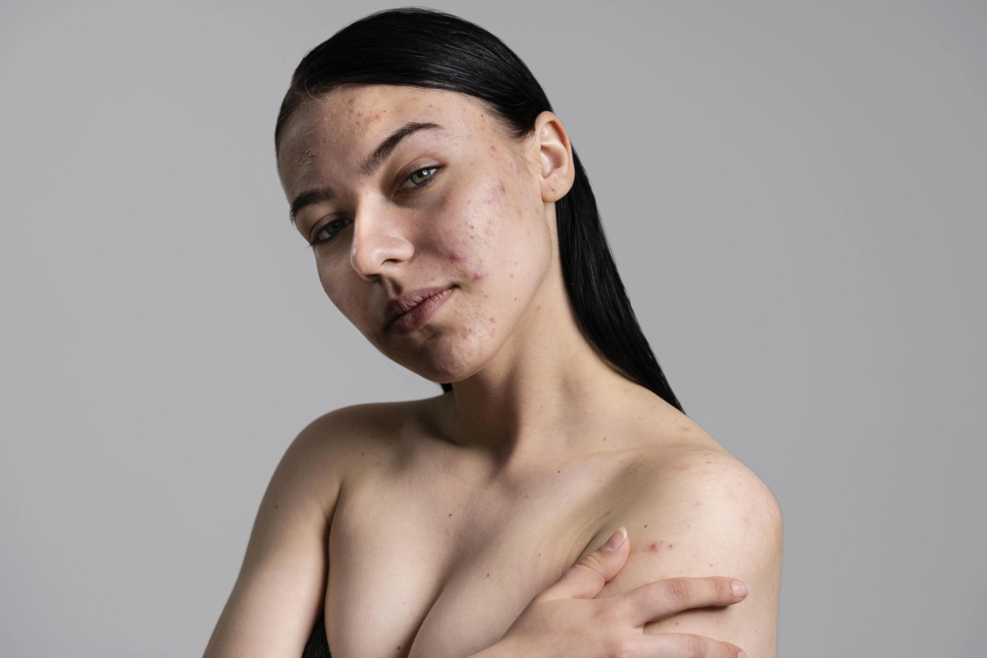
Appearance

Seborrheic keratoses can have various appearances:
• Shape — they can be flat or raised nodules.
• Surface — often scaly, sometimes fissured.
• Color — ranges from skin-colored to yellowish, brown, or black.
• Size — from 1 mm to several centimeters.
• Location — almost anywhere on the body, except the palms and soles.
• They often appear in clusters, especially on the chest, back, scalp, and in skin folds.
On the back, they sometimes arrange themselves in a "Christmas tree-like" pattern, which is related to their spread along the lines of Blaschko.

Complications

A seborrheic keratosis is a benign growth and is not cancer or a precancerous condition. However, in some cases, its appearance can mimic malignant growths, such as:
• Basal cell carcinoma.
• Squamous cell carcinoma.
• Melanoma.
Additionally, the sudden and massive eruption of seborrheic keratoses (especially in older individuals) can be a sign of an internal malignancy, such as gastric adenocarcinoma. This phenomenon is known as the
sign of Leser-Trélat.
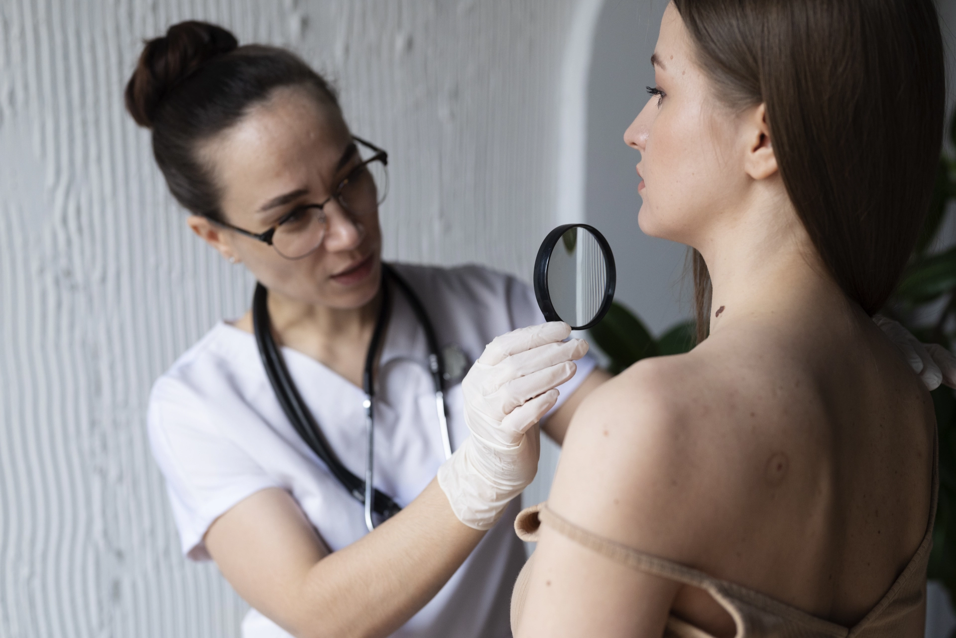
Diagnosis

1. Clinical examination — in many cases, a diagnosis can be made based on a visual inspection.
2. Dermoscopy — a non-invasive method that helps differentiate seborrheic keratosis from malignant growths. Characteristic dermoscopic features include:
• Comedo-like openings.
• Milia-like cysts.
• Gyri and sulci ("brain-like" structure).
• Hairpin-like vessels.
3. Biopsy and histological examination — performed in suspicious cases where dermoscopic or clinical data are not conclusive.

Differential Diagnosis

Seborrheic keratosis must be differentiated from the following growths:
• Common warts (verruca vulgaris).
• Actinic keratosis.
• Pigmented basal cell carcinoma.
• Squamous cell carcinoma.
• Melanoma — rare, but important to rule out.

Treatment

Seborrheic keratosis doesn't require treatment, but it can be removed in the following situations:
• Aesthetic discomfort.
• Itching or irritation.
• Injury from clothing or shaving.

Treatment methods:

• Cryodestruction (freezing).
• Electrocoagulation.
• Laser therapy.
• Shave biopsy.
The choice of treatment method is based on the size and location of the growth, as well as the patient's overall health. When choosing the best method, all indications and contraindications should be considered.

Prevention and Prognosis

Since the exact causes of its occurrence are not fully understood, there are no specific preventive measures known. However, it is recommended to:
• Use sunscreen.
• Undergo regular dermatological examinations.
• Avoid excessive sun exposure.
Some seborrheic keratoses can disappear on their own over time. In suspicious cases, you should always consult a specialist.
Acute and Chronic Pancreatitis: What you need to know
05 September 2025
The pancreas is a small but extremely important organ responsible for digestion and insulin production. When it becomes inflamed, it leads to pancreatitis, a disease that can cause serious complications if left untreated. Acute Pancreatitis Acute pancreatitis starts suddenly and is usually accompanied by very severe pain in the upper abdomen. It's often linked to alcohol consumption or excessively fatty foods. Symptoms: • Sharp and unbearable abdominal pain • Nausea and vomiting • High fever • General weakness This is a dangerous condition that requires immediate medical intervention. Sometimes hospitalization is necessary. The disease may appear once and be cured with treatment, but in some cases, it can recur and become chronic. Treatment: In cases of acute pancreatitis, the patient is usually admitted to the hospital. For the first 1-2 days, no food is allowed to give the pancreas a "rest". Fluids and nutrients are provided intravenously. Pain relievers, anti-inflammatory drugs, and sometimes antibiotics are used. Infusion therapy (serums, electrolytes) is prescribed to maintain the body's balance. The diet starts with liquids, and then dietary foods are gradually added. In severe cases, surgery may be required. Chronic Pancreatitis The chronic form is a long-term disease that develops over years. The main causes are: • Long-term alcohol use • Dietary violations • Frequent recurring acute pancreatitis Symptoms: The pancreas is gradually damaged. The symptoms are: • Constant or recurring abdominal pain • Bloating • Digestive disorders, diarrhea • Unexplained weight loss Over time, diabetes may develop as the pancreas stops producing enough insulin. Treatment: Chronic pancreatitis requires long-term management. The diet is strictly important: • Alcohol, fatty, fried, and spicy foods must be excluded. • It's recommended to eat small portions, but frequently. • Enzyme preparations are used to aid digestion. • Pain relievers are used for pain. • If diabetes develops, insulin or other blood sugar-regulating drugs are prescribed. Don't forget that early diagnosis and treatment prevent the exacerbation and progression of the disease. If you have symptoms of pancreatitis, consult a specialist immediately. Contact the Dalimed medical center to get a proper diagnosis and appropriate treatment.
Rosacea
03 September 2025
Rosacea is a chronic inflammatory skin disease that mainly affects the central part of the face. It is characterized by persistent redness, the appearance of pustules and nodules, and also hypersensitivity of the skin. The disease is most common in people with fair skin and is more prevalent among women, but in men the disease can be more severe. Causes and mechanism of development The exact cause of rosacea is still not fully understood. It is believed that several factors play a role in the development of the disease: • Vascular dysfunction: superficial vessels of the face easily dilate, which leads to redness. • Immune system hypersensitivity: the skin reacts too strongly to external irritants. • Microorganisms: the Demodex folliculorum mite and certain bacteria can contribute to an inflammatory reaction. • Genetic predisposition: the disease is more common in some families. Main clinical forms Rosacea manifests in various forms: • Erythematotelangiectatic form – characterized by persistent facial redness and a network of small vessels. • Papulopustular form – pustules and nodules appear on the face, which are often confused with acne. • Phymatous form – the skin thickens, most often in the nose area, leading to rhinophyma. Aggravating factors The course of rosacea is characterized by phases of flare-ups and remission. Flare-ups often occur due to the following factors: • Sun exposure • Hot or very cold weather • Stress • Alcoholic beverages • Spicy food, hot drinks • Irritating ingredients in cosmetic products Diagnosis The diagnosis of rosacea is carried out mainly on the basis of the clinical picture. Principles of treatment Rosacea treatment is individual and depends on the form and severity of the disease. • Topical treatment: creams or gels with metronidazole, azelaic acid, or ivermectin. • Systemic treatment: a long course of doxycycline or other tetracycline antibiotics. • Laser therapy: useful for reducing vascular networks and redness. • Surgical intervention: in the case of severe rhinophyma, surgical correction may be applied. Care and prevention In addition to medical treatment, proper skin care is of great importance: • Use gentle, soap-free cleansing products • Apply sunscreen with SPF 50+ • Avoid aggravating factors • Moisturize the skin with non-irritating products Conclusion Rosacea is a chronic but controllable disease. Although there is no complete cure, timely diagnosis and proper treatment can significantly reduce symptoms and improve quality of life. Patients are advised to regularly visit a dermatologist and follow the rules of care.
Anti-Müllerian Hormone
01 September 2025
Anti-Müllerian hormone (AMH) is a protein substance that is produced in the body of both men and women. Main functions of AMH In women, AMH is synthesized by cells in the ovaries and reflects the quantity and quality of eggs, meaning it is an important indicator of reproductive health. In women, the level of anti-Müllerian hormone shows: • ovarian reserve—the number of maturing follicles and potential eggs. • the probability of successful conception naturally or with IVF. • the onset of fertility decline (AMH levels decrease with age). • possible disorders, for example, polycystic ovary syndrome (PCOS), in which the hormone level is higher than normal. In men, the hormone plays a role in the formation of the reproductive system during the prenatal period. In men, AMH is responsible for the proper formation of the reproductive system in the fetus and is used in the diagnosis of certain developmental and reproductive disorders. When the analysis is prescribed The anti-Müllerian hormone test is recommended in the following cases: • Fertility assessment and pregnancy planning. • Selection and monitoring of assisted reproductive technology programs (for example, IVF). • Suspicion of premature ovarian failure or PCOS. • Evaluation of ovarian function after surgery, chemo-, or radiation therapy. • Diagnosis of sexual development disorders in boys. Normal and abnormal levels A high level of AMH may indicate PCOS or delayed puberty. A low level of AMH is most often associated with a decrease in ovarian reserve, the onset of menopause, or premature ovarian failure. It should be taken into account that the results of the analysis are always interpreted in conjunction with other studies and the clinical picture. How the study is conducted Blood is taken from a vein for the analysis. No special preparation is required, but it is recommended to take the test in the morning and on an empty stomach. Why it's important to take an AMH test Anti-Müllerian hormone is one of the key markers of female reproductive health. Timely examination helps to correctly plan a pregnancy, monitor IVF programs, and identify possible disorders in the ovaries. The AMH analysis can be submitted at the Dalimed medical center, where modern diagnostic methods are used and specialists are ready to help you preserve your health and realize your reproductive plans.
Appendicitis
29 August 2025
Appendicitis is an inflammation of the appendix, a small pouch attached to the large intestine. Despite its tiny size, the condition can be extremely dangerous: an inflamed appendix quickly swells and, if it ruptures, can cause a severe infection of the abdominal cavity known as peritonitis. For this reason, appendicitis is considered a medical emergency and requires immediate medical attention.CausesThe most common cause is an obstruction of the appendix's lumen by fecal matter. Less frequently, inflammation can be caused by an infection, parasites, or a tumor. This obstruction leads to bacterial growth, swelling, and impaired blood flow to the tissues. Without treatment, the appendix wall gradually deteriorates and can rupture.FormsAcute appendicitis develops suddenly, is accompanied by severe pain, and progresses rapidly.Chronic appendicitis is rare and manifests as intermittent pain. However, it can turn into the acute form at any time, so it requires a doctor's supervision.SymptomsThe most characteristic signs include:• Pain that starts near the navel and then moves to the lower right abdomen, intensifying with movement or coughing.• Nausea, vomiting, and loss of appetite.• Low-grade fever, sometimes with chills.• Changes in bowel habits: constipation, diarrhea, or difficulty passing gas.• Abdominal bloating and tension.It's important to note that the classic symptoms are not always present in children, the elderly, and pregnant women. Therefore, any sudden abdominal discomfort warrants an urgent consultation.DiagnosisTo confirm the diagnosis, a doctor will perform an examination and order blood and urine tests. Additionally, imaging methods are used:• Ultrasound helps to detect inflammation.• CT scan provides a detailed picture.• MRI is more often used in pregnant women to reduce radiation exposure.TreatmentAppendicitis is treated surgically. The main methods are:• Classic appendectomy, performed through an incision in the lower abdomen.• Laparoscopy, a minimally invasive surgery using small incisions and a camera.For complicated cases, drainage of the abdominal cavity and a course of antibiotics may be necessary. Sometimes, antibiotics are used as a temporary measure before surgery, but they cannot fully replace it.ComplicationsThe main threat is the rupture of the appendix and the development of peritonitis. In this case, the infection spreads throughout the abdominal cavity and can lead to sepsis, a life-threatening condition. Without treatment, the risk of a fatal outcome is very high.Appendicitis is the most common surgical disease in people aged 10–30, but it can occur at any age. Its danger lies in the fact that only 48–72 hours can pass from the first symptoms to a rupture. When you experience sudden abdominal pain, do not delay a visit to the doctor. Timely medical attention is the guarantee of successful treatment and good health.

Fill in the required fields

I am waiting for a call

We will help you quickly find what you need!

Thank you!
Our employee will contact you soon
Close
dalimed medical
How can we help?
Leave your number and we will call you back
Book a call
I agree with the terms of personal data processing
Thank you!
Our employee will contact you soon
Close