Jaundice in Newborns

Jaundice in Newborns

Jaundice in Newborns
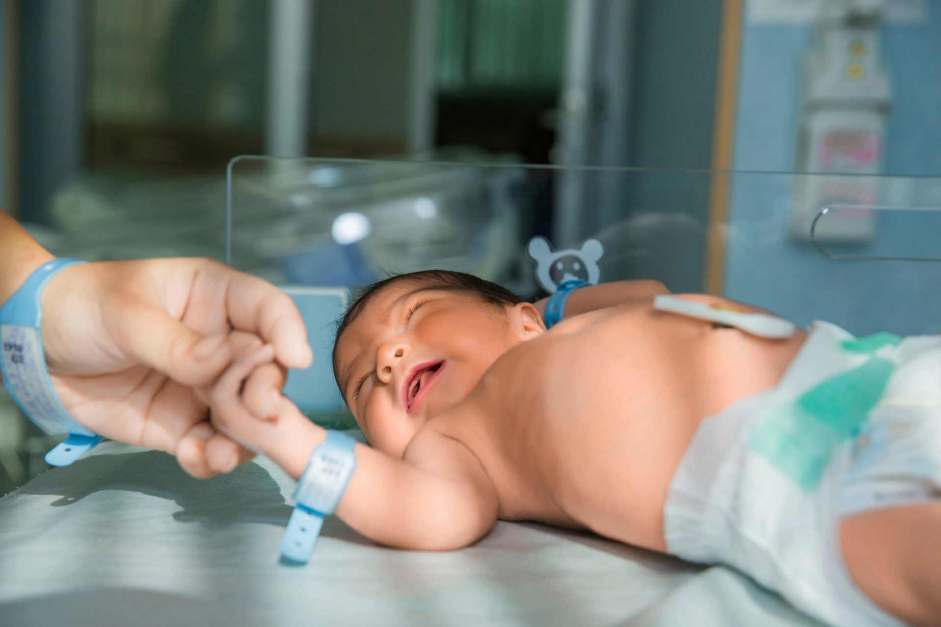
It’s one of the first things new parents might notice after birth — a yellowish tint to their baby’s skin or eyes. This condition, known as jaundice, is extremely common in newborns. In fact, it affects up to 60% of full-term babies and as many as 80% of preterm infants in the first week of life.
While it’s usually harmless and temporary, understanding why jaundice happens — and when it might signal a more serious issue — is important for every parent.

What Is Jaundice and Why Does It Happen?

Jaundice in newborns occurs when there’s too much bilirubin in the baby’s blood — a condition called hyperbilirubinemia. Bilirubin is a yellow pigment produced during the normal breakdown of red blood cells.
Before birth, the mother's liver does all the work of removing bilirubin for the baby. But after birth, that job shifts to the baby’s own liver — which may not be fully developed yet. If the liver can’t filter out bilirubin quickly enough, the substance starts to build up in the baby’s body, giving their skin and eyes a yellow hue.
This yellowing typically starts on the face and can spread to the chest, abdomen, arms, and legs as bilirubin levels rise. It’s easiest to spot in natural light, and in babies with darker skin, it may be more noticeable in the whites of the eyes or under the tongue.

Types of Newborn Jaundice

Not all jaundice is the same. There are several types, each with different causes and timeframes:
1.

Physiological Jaundice

This is the most common type and occurs in nearly all newborns to some extent. It usually appears within 2 to 3 days after birth and disappears on its own within one to two weeks. It’s a natural part of the baby’s adjustment to life outside the womb as their liver matures.
2.

Breastfeeding Jaundice

This can develop in the first week of life when a baby isn’t getting enough breast milk. It may be due to difficulties with latching, a delay in milk coming in, or infrequent feedings. Less feeding means fewer bowel movements — and that leads to slower elimination of bilirubin from the body.
3.

Breast Milk Jaundice

This is different from breastfeeding jaundice. It typically appears after the first week and can last for a month or longer. In this case, certain substances in the mother's milk may interfere with how the baby’s liver processes bilirubin. Although it sounds concerning, breast milk jaundice is usually harmless and doesn’t require stopping breastfeeding.
4.

Jaundice from Medical Conditions

In some rare cases, jaundice can be caused by underlying health problems, such as:
•Blood type incompatibility between mother and baby
•Bruising during birth (which increases red blood cell breakdown)
•Infections like sepsis
•Liver conditions, including biliary atresia
•Low oxygen levels at birth
•An excess of red blood cells
These forms of jaundice are less common but more serious and require immediate medical attention.

Symptoms to Watch For

The most obvious sign is yellowing of the skin and eyes. But parents should also monitor for other warning signs that may indicate severe jaundice or related complications:
•Bright yellow or orange-tinted skin
•Excessive sleepiness or trouble waking for feeds
•Baby is very fussy or difficult to calm
•Poor feeding, whether at the breast or with a bottle
•Not enough wet or dirty diapers
If jaundice seems to be getting worse after the first few days — or lasts longer than two weeks — it’s time to call your pediatrician.

How Is Jaundice Treated?

In most cases, no treatment is needed. As your baby’s liver matures and they begin feeding regularly, bilirubin levels naturally decrease. Frequent feedings — around 10 to 12 times a day — help stimulate more bowel movements, which remove bilirubin through the stool.
However, if bilirubin levels are too high or rising quickly, your baby’s doctor may recommend phototherapy — a safe and effective treatment that uses special blue lights to help break down bilirubin in the skin.
In rare, severe cases, where phototherapy isn’t enough, a treatment called an exchange transfusion may be necessary. This involves replacing a portion of the baby’s blood with donor blood to quickly reduce bilirubin levels. Fortunately, this is rarely needed with early monitoring and intervention.
While jaundice in newborns can be unsettling for new parents, it’s important to know that it’s usually a normal part of development. Most cases resolve without complications, especially when babies are well-fed and monitored closely.
Still, severe jaundice can pose serious risks, including brain damage if left untreated — so staying informed and knowing when to call your healthcare provider is essential.
If you’re ever unsure, don’t hesitate to reach out to your pediatrician. When caught early, jaundice is almost always treatable and temporary.
What is glycated hemoglobin?
20 April 2026
Glycated hemoglobin, commonly known as HbA1c or simply A1C, is a key laboratory marker used to evaluate average blood sugar levels over time. Unlike a single blood glucose measurement that reflects only a moment in time, HbA1c provides a broader picture of how glucose has behaved in the body over the past two to three months. This makes it an essential tool in the detection and management of diabetes. How does HbA1c form? HbA1c is created through a natural process called glycation. When glucose circulates in the bloodstream, a portion of it attaches to hemoglobin — the protein inside red blood cells responsible for carrying oxygen. This binding happens gradually and continuously throughout the lifespan of red blood cells, which is about 120 days. The process occurs in two main stages: • First, glucose temporarily binds to hemoglobin, forming an unstable intermediate structure. • Then, this structure undergoes a chemical transformation into a stable compound known as glycated hemoglobin. Because this attachment is irreversible, the amount of HbA1c reflects the average level of glucose exposure over time. More glucose in the blood leads to higher HbA1c values. What does the HbA1c test measure? The HbA1c test determines the percentage of hemoglobin that has glucose attached to it. The result is expressed as a percentage, which corresponds to average blood sugar levels over the previous weeks. Typical interpretation includes: • Below 5.7% — normal range • 5.7% to 6.4% — increased risk (prediabetes) • 6.5% or higher — consistent with diabetes Healthcare providers use this test not only for diagnosis but also to monitor how well blood sugar is controlled over time. Why is HbA1c important? HbA1c plays a central role in long-term diabetes care because it offers insights that daily glucose checks cannot fully capture. It helps both patients and healthcare providers understand overall trends rather than isolated readings. Key benefits include: • Long-term assessment: Reflects average glucose levels over 2–3 months • Convenience: Does not require fasting and can be done at any time of day • Treatment guidance: Helps adjust medications, diet, and lifestyle strategies • Risk evaluation: Higher levels are associated with complications such as cardiovascular disease, nerve damage, kidney problems, and vision impairment Maintaining HbA1c within the target range significantly reduces the risk of these complications. How often should HbA1c be tested? For individuals diagnosed with diabetes, HbA1c is usually checked at least twice a year. More frequent testing may be needed if treatment changes or if blood sugar levels are not well controlled. It may also be used as a screening test in people with risk factors such as obesity, family history of diabetes, or low physical activity. Limitations of the HbA1c test Although HbA1c is highly useful, certain conditions can affect its accuracy. These include: • Disorders that shorten or prolong red blood cell lifespan (such as anemia or recent blood loss) • Hemoglobin variants (genetic differences in hemoglobin structure) • Pregnancy, especially in later stages • Advanced kidney disease In such cases, HbA1c results should be interpreted carefully and may need to be combined with other tests. Conclusion Glycated hemoglobin (HbA1c) is a reliable and widely used indicator of long-term blood sugar control. By reflecting average glucose levels over several months, it provides valuable guidance for diagnosing diabetes, monitoring treatment effectiveness, and reducing the risk of complications.
Dental plaque removal
15 April 2026
Dental plaque is a soft, sticky film that continuously forms on teeth as a result of normal biological processes in the mouth. It consists of bacteria, saliva components, and remnants of food. Although it is a natural occurrence, plaque becomes harmful when it is allowed to accumulate. Shortly after cleaning your teeth, a thin invisible layer forms on the enamel. This layer attracts bacteria, which begin to multiply rapidly. When food is consumed—especially sugars and starches—these bacteria feed on the particles and produce acids. This combination of bacteria, acids, and food debris forms plaque. Because this process happens constantly, plaque begins to rebuild within hours after brushing. What happens if plaque is not removed Plaque is the starting point of most common dental problems. If left untreated, it can lead to: • Tooth decay: Acids break down enamel, forming cavities • Gingivitis: Early gum disease characterized by redness, swelling, and bleeding • Periodontal disease: Advanced infection affecting supporting bone and tissues • Tartar formation: Hardened plaque that cannot be removed at home Additionally, plaque buildup is a common cause of persistent bad breath due to bacterial activity. Daily plaque removal strategies Effective plaque control requires a combination of habits: • Brushing twice daily: Use gentle, thorough movements and clean all tooth surfaces • Cleaning between teeth: Floss or use interdental tools to remove hidden plaque • Supporting products: Antibacterial or fluoride mouthwash may enhance protection Consistency is key, as plaque reforms quickly. Professional plaque and tartar removal When plaque hardens into tartar, it firmly attaches to teeth and often accumulates along the gum line. At this stage, only dental professionals can remove it through a procedure known as scaling. Professional cleanings may also include polishing, which smooths the tooth surface and helps reduce future plaque buildup. Why regular dental visits matter Routine dental check-ups allow early detection of problems caused by plaque. Depending on individual risk factors, visits are typically recommended every six months, though some individuals may require more frequent care. Conclusion Dental plaque is a natural but potentially harmful part of oral biology. Without proper care, it can lead to a range of complications affecting both teeth and gums. However, with consistent daily hygiene and regular professional care, plaque can be effectively controlled. Schedule your routine check-up today to ensure a healthy smile and prevent future complications.
What is hyperprolactinemia?
13 April 2026
Hyperprolactinemia is a condition characterized by abnormally high levels of prolactin, a hormone secreted by the pituitary gland. While prolactin is essential for breastfeeding, it also interacts with other hormones that regulate reproductive function. When its levels become elevated outside of pregnancy, it can disrupt the endocrine system and affect multiple processes in the body. One of the key effects of excess prolactin is the suppression of reproductive hormones. In women, this leads to decreased estrogen levels, which can interfere with ovulation and menstrual regularity. Over time, these hormonal changes can also influence bone metabolism, increasing the risk of bone loss. People with hyperprolactinemia may experience a variety of symptoms, although some remain asymptomatic. The most typical manifestations include: • irregular or absent menstrual cycles and infertility • decreased libido and sexual dysfunction • unexpected milk discharge (galactorrhea) The condition has several possible causes. The most common is a benign tumor of the pituitary gland known as a prolactinoma, which produces excess prolactin. Other contributing factors include medications that interfere with dopamine regulation, thyroid dysfunction, chronic diseases, or physical stress affecting the chest wall. In a number of cases, no specific cause can be identified. Diagnosis is usually simple and begins with measuring prolactin levels in the blood. If elevated levels are confirmed, further evaluation such as MRI imaging may be recommended to determine the underlying cause. Treatment is guided by the underlying cause but is usually highly effective. Most patients are treated with medications that help reduce prolactin levels and restore normal hormonal balance. In less common cases, surgical or other specialized treatments may be necessary. With proper management, the outlook is generally excellent, and most people experience significant improvement in symptoms.
Ultrasound at 12 week of pregnancy
09 April 2026
The 12-week pregnancy scan is one of the most important early prenatal examinations because it provides the first detailed medical assessment of how the pregnancy is developing. Performed between 11 weeks and 13 weeks + 6 days, this ultrasound helps confirm that the pregnancy is progressing normally, establish accurate gestational age, and detect early findings that may influence further care. For many women, it is also the first clear view of the baby, but its main purpose is medical evaluation and safe pregnancy monitoring. Why the scan is needed This examination helps doctors: • confirm fetal heartbeat • calculate the due date accurately • determine whether there is one baby or multiple pregnancy • confirm that the pregnancy is developing inside the uterus Early dating at this stage is considered highly reliable for planning the rest of pregnancy care. What doctors check during the examination At 12 weeks, several important fetal structures are already visible. The scan evaluates: • head and spine • arms and legs • stomach and bladder • placenta and umbilical cord The uterus and ovaries are also checked because maternal findings may affect follow-up. Nuchal translucency measurement A key part of the scan is measuring nuchal translucency (NT), the fluid behind the baby’s neck. This helps estimate the risk of chromosomal conditions such as Down syndrome, Edwards syndrome, and Patau syndrome. When combined with blood tests, this screening helps decide whether additional investigations are needed. Why early detection is important The scan can reveal early structural concerns, placental findings, or pregnancy risks that may require closer monitoring later. It also provides reassurance when fetal heartbeat and growth appear normal. For accurate early pregnancy assessment, you can have this examination at Dalimed medical center, where experienced specialists provide careful prenatal ultrasound evaluation.

Fill in the required fields

I am waiting for a call

We will help you quickly find what you need!

Thank you!
Our employee will contact you soon
Close
dalimed medical
How can we help?
Leave your number and we will call you back
Book a call
I agree with the terms of personal data processing
Thank you!
Our employee will contact you soon
Close