What to do when the baby has an earache?

What to do when the baby has an earache?

What to do when the baby has an earache?
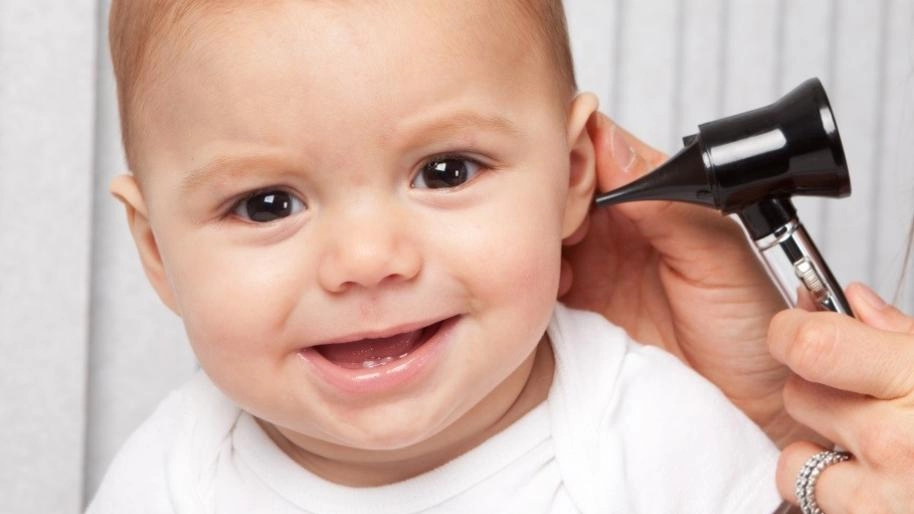
Earache is common among children, especially in early childhood. The presence of earache usually means that the child has an ear infection, otitis. Almost all parents deal with ear infections in children. This disease is especially typical for children aged 1-3, but it can also occur at a younger age. According to epidemiological studies, all children up to 5-7 years old get sick with this disease at least once, especially often at the age of 6-12 months. And after that age, from 5-7 years old, ear infections rarely occur. This is due to the structural features of the auditory tube in children.
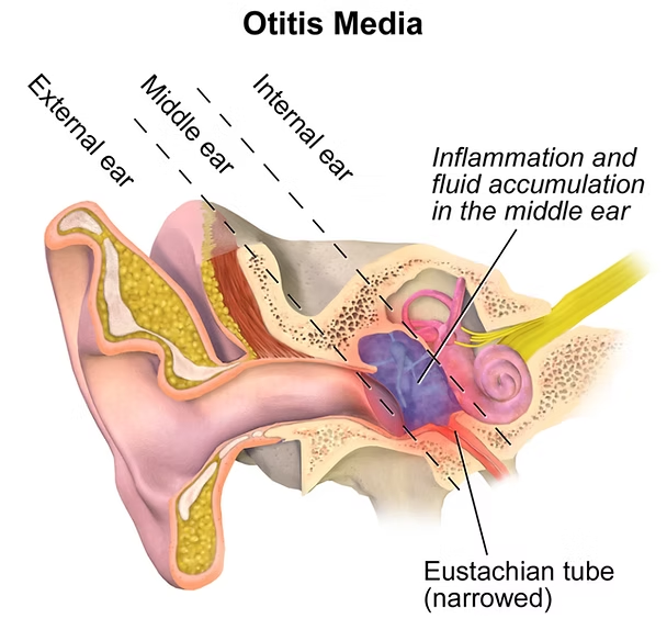
There are external, middle and internal ear infections. External ear infections occur when the infection accumulates around the hairs of the auditory tube, which can also cause an abscess.
Otitis media is a complication of a viral or bacterial infection, or it can occur as a result of a ruptured eardrum. Inflammation of the inner ear occurs as a result of an infection of the paranasal sinuses, inflammation of the meninges, or purulent middle ear inflammation. Otitis media is more common in children.

What are the causes of earache in children?

Viral and bacterial infections

, in which the space behind the eardrum becomes inflamed and the middle ear becomes inflamed.

Swimmer's ear

, which occurs as a result of irritation of the external auditory canal by water. The main symptom is itching in the auditory canal. If there is an infection, ear pain also occurs.

Injury to the auditory canal

, which occurs when a hard object is used to clean the ear. The abrasions can become inflamed, which contributes to the occurrence of pain.

An abscess

in the ear canal, which can be very painful.

A wax plug

, which is a solid lump of earwax. It causes mild pain, but if the lump goes deeper into the ear canal when you clean the ear with a cotton swab, it can block the ear canal, cause inflammation, pain, and hearing loss in that ear.

Foreign bodies

. Children can put various objects in their ears that can cause pain. These are usually small objects found in toys. Do not let your child play with small toys that are not recommended for children of their age, especially without parental supervision.

Barotrauma

. During pressure fluctuations, such as on an airplane or in the elevators of high-rise buildings, the eardrum can stretch, which can cause pain, especially when there is a wax plug in the ear.

Referred pain

. Earache can be caused by other problems, such as inflammation of the tonsils, jaw, parotid gland, mandibular glands, or teething.

What to do when the baby has an earache?

The main signs of ear inflammation are fever, general weakness, restlessness, putting a hand to the ear, crying, ear pain or a feeling of blockage in the ear, decreased hearing, and discharge from the ear.

Sometimes a child may not have earache, but have a high fever and general weakness, nausea, and ear inflammation is confirmed during the examination. This situation is especially common in children under 3 years of age.
The following signs indicate the presence of an external ear infection: fever and ear pain, which increases when chewing and talking.
With inflammation of the middle ear, the child complains of sharp pain in the ear, impaired hearing, and sometimes noise in the ear. There may also be pus production and a fever of up to 38-40 ° С. Breast-fed children may refuse to breastfeed and reach for the sore ear.
Inflammation of the inner ear is characterized by a sharp decrease in hearing, balance disorders, headache, and nausea.
In the absence of treatment, the process can worsen and lead to complications and hearing loss.
Therefore, if you notice such signs in your child, do not self-medicate, immediately contact our clinic, and our pediatricians will quickly diagnose whether the child has otitis media by examining the ear, and if necessary, refer them to a specialist.
In our clinic, children are treated according to evidence-based principles of medicine: we select only those diagnostic and treatment methods that have proven their effectiveness and never prescribe unnecessary examinations and medications to fully maintain your child’s health.

What is glycated hemoglobin?
20 April 2026
Glycated hemoglobin, commonly known as HbA1c or simply A1C, is a key laboratory marker used to evaluate average blood sugar levels over time. Unlike a single blood glucose measurement that reflects only a moment in time, HbA1c provides a broader picture of how glucose has behaved in the body over the past two to three months. This makes it an essential tool in the detection and management of diabetes. How does HbA1c form? HbA1c is created through a natural process called glycation. When glucose circulates in the bloodstream, a portion of it attaches to hemoglobin — the protein inside red blood cells responsible for carrying oxygen. This binding happens gradually and continuously throughout the lifespan of red blood cells, which is about 120 days. The process occurs in two main stages: • First, glucose temporarily binds to hemoglobin, forming an unstable intermediate structure. • Then, this structure undergoes a chemical transformation into a stable compound known as glycated hemoglobin. Because this attachment is irreversible, the amount of HbA1c reflects the average level of glucose exposure over time. More glucose in the blood leads to higher HbA1c values. What does the HbA1c test measure? The HbA1c test determines the percentage of hemoglobin that has glucose attached to it. The result is expressed as a percentage, which corresponds to average blood sugar levels over the previous weeks. Typical interpretation includes: • Below 5.7% — normal range • 5.7% to 6.4% — increased risk (prediabetes) • 6.5% or higher — consistent with diabetes Healthcare providers use this test not only for diagnosis but also to monitor how well blood sugar is controlled over time. Why is HbA1c important? HbA1c plays a central role in long-term diabetes care because it offers insights that daily glucose checks cannot fully capture. It helps both patients and healthcare providers understand overall trends rather than isolated readings. Key benefits include: • Long-term assessment: Reflects average glucose levels over 2–3 months • Convenience: Does not require fasting and can be done at any time of day • Treatment guidance: Helps adjust medications, diet, and lifestyle strategies • Risk evaluation: Higher levels are associated with complications such as cardiovascular disease, nerve damage, kidney problems, and vision impairment Maintaining HbA1c within the target range significantly reduces the risk of these complications. How often should HbA1c be tested? For individuals diagnosed with diabetes, HbA1c is usually checked at least twice a year. More frequent testing may be needed if treatment changes or if blood sugar levels are not well controlled. It may also be used as a screening test in people with risk factors such as obesity, family history of diabetes, or low physical activity. Limitations of the HbA1c test Although HbA1c is highly useful, certain conditions can affect its accuracy. These include: • Disorders that shorten or prolong red blood cell lifespan (such as anemia or recent blood loss) • Hemoglobin variants (genetic differences in hemoglobin structure) • Pregnancy, especially in later stages • Advanced kidney disease In such cases, HbA1c results should be interpreted carefully and may need to be combined with other tests. Conclusion Glycated hemoglobin (HbA1c) is a reliable and widely used indicator of long-term blood sugar control. By reflecting average glucose levels over several months, it provides valuable guidance for diagnosing diabetes, monitoring treatment effectiveness, and reducing the risk of complications.
Dental plaque removal
15 April 2026
Dental plaque is a soft, sticky film that continuously forms on teeth as a result of normal biological processes in the mouth. It consists of bacteria, saliva components, and remnants of food. Although it is a natural occurrence, plaque becomes harmful when it is allowed to accumulate. Shortly after cleaning your teeth, a thin invisible layer forms on the enamel. This layer attracts bacteria, which begin to multiply rapidly. When food is consumed—especially sugars and starches—these bacteria feed on the particles and produce acids. This combination of bacteria, acids, and food debris forms plaque. Because this process happens constantly, plaque begins to rebuild within hours after brushing. What happens if plaque is not removed Plaque is the starting point of most common dental problems. If left untreated, it can lead to: • Tooth decay: Acids break down enamel, forming cavities • Gingivitis: Early gum disease characterized by redness, swelling, and bleeding • Periodontal disease: Advanced infection affecting supporting bone and tissues • Tartar formation: Hardened plaque that cannot be removed at home Additionally, plaque buildup is a common cause of persistent bad breath due to bacterial activity. Daily plaque removal strategies Effective plaque control requires a combination of habits: • Brushing twice daily: Use gentle, thorough movements and clean all tooth surfaces • Cleaning between teeth: Floss or use interdental tools to remove hidden plaque • Supporting products: Antibacterial or fluoride mouthwash may enhance protection Consistency is key, as plaque reforms quickly. Professional plaque and tartar removal When plaque hardens into tartar, it firmly attaches to teeth and often accumulates along the gum line. At this stage, only dental professionals can remove it through a procedure known as scaling. Professional cleanings may also include polishing, which smooths the tooth surface and helps reduce future plaque buildup. Why regular dental visits matter Routine dental check-ups allow early detection of problems caused by plaque. Depending on individual risk factors, visits are typically recommended every six months, though some individuals may require more frequent care. Conclusion Dental plaque is a natural but potentially harmful part of oral biology. Without proper care, it can lead to a range of complications affecting both teeth and gums. However, with consistent daily hygiene and regular professional care, plaque can be effectively controlled. Schedule your routine check-up today to ensure a healthy smile and prevent future complications.
What is hyperprolactinemia?
13 April 2026
Hyperprolactinemia is a condition characterized by abnormally high levels of prolactin, a hormone secreted by the pituitary gland. While prolactin is essential for breastfeeding, it also interacts with other hormones that regulate reproductive function. When its levels become elevated outside of pregnancy, it can disrupt the endocrine system and affect multiple processes in the body. One of the key effects of excess prolactin is the suppression of reproductive hormones. In women, this leads to decreased estrogen levels, which can interfere with ovulation and menstrual regularity. Over time, these hormonal changes can also influence bone metabolism, increasing the risk of bone loss. People with hyperprolactinemia may experience a variety of symptoms, although some remain asymptomatic. The most typical manifestations include: • irregular or absent menstrual cycles and infertility • decreased libido and sexual dysfunction • unexpected milk discharge (galactorrhea) The condition has several possible causes. The most common is a benign tumor of the pituitary gland known as a prolactinoma, which produces excess prolactin. Other contributing factors include medications that interfere with dopamine regulation, thyroid dysfunction, chronic diseases, or physical stress affecting the chest wall. In a number of cases, no specific cause can be identified. Diagnosis is usually simple and begins with measuring prolactin levels in the blood. If elevated levels are confirmed, further evaluation such as MRI imaging may be recommended to determine the underlying cause. Treatment is guided by the underlying cause but is usually highly effective. Most patients are treated with medications that help reduce prolactin levels and restore normal hormonal balance. In less common cases, surgical or other specialized treatments may be necessary. With proper management, the outlook is generally excellent, and most people experience significant improvement in symptoms.
Ultrasound at 12 week of pregnancy
09 April 2026
The 12-week pregnancy scan is one of the most important early prenatal examinations because it provides the first detailed medical assessment of how the pregnancy is developing. Performed between 11 weeks and 13 weeks + 6 days, this ultrasound helps confirm that the pregnancy is progressing normally, establish accurate gestational age, and detect early findings that may influence further care. For many women, it is also the first clear view of the baby, but its main purpose is medical evaluation and safe pregnancy monitoring. Why the scan is needed This examination helps doctors: • confirm fetal heartbeat • calculate the due date accurately • determine whether there is one baby or multiple pregnancy • confirm that the pregnancy is developing inside the uterus Early dating at this stage is considered highly reliable for planning the rest of pregnancy care. What doctors check during the examination At 12 weeks, several important fetal structures are already visible. The scan evaluates: • head and spine • arms and legs • stomach and bladder • placenta and umbilical cord The uterus and ovaries are also checked because maternal findings may affect follow-up. Nuchal translucency measurement A key part of the scan is measuring nuchal translucency (NT), the fluid behind the baby’s neck. This helps estimate the risk of chromosomal conditions such as Down syndrome, Edwards syndrome, and Patau syndrome. When combined with blood tests, this screening helps decide whether additional investigations are needed. Why early detection is important The scan can reveal early structural concerns, placental findings, or pregnancy risks that may require closer monitoring later. It also provides reassurance when fetal heartbeat and growth appear normal. For accurate early pregnancy assessment, you can have this examination at Dalimed medical center, where experienced specialists provide careful prenatal ultrasound evaluation.

Fill in the required fields

I am waiting for a call

We will help you quickly find what you need!

Thank you!
Our employee will contact you soon
Close
dalimed medical
How can we help?
Leave your number and we will call you back
Book a call
I agree with the terms of personal data processing
Thank you!
Our employee will contact you soon
Close