Knee Joint Pain

Knee Joint Pain

Knee Joint Pain
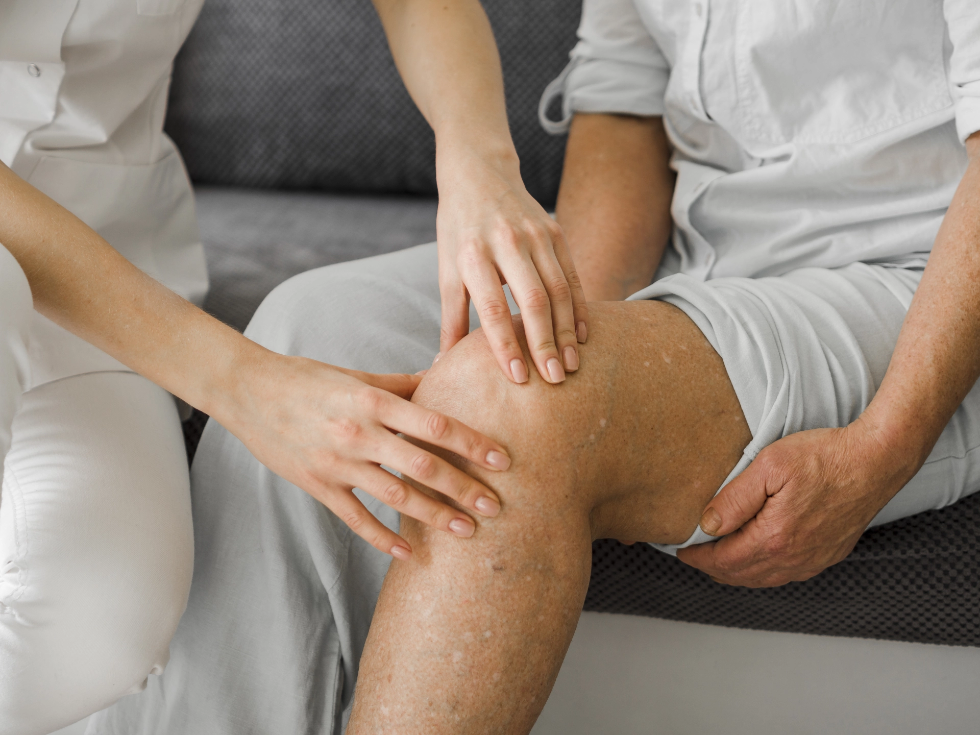
The knee joint is subjected to heavy daily stress. It is a fairly large joint, surrounded inside and outside by ligaments and tendons that ensure its stability. In fact, the articular cartilage itself has no nerve supply. With age, as cartilage wears down and body weight increases, the strain on the ligamentous apparatus also increases, leading to inflammation and destructive joint processes. At this stage, the patient feels pain in the knee joint.
According to global statistics, every 10th person over the age of 55 suffers from gonarthritis, and one in four of them becomes disabled and requires knee joint endoprosthesis surgery.

Causes of knee pain:

• Osteoarthritis, mainly caused by cartilage wear,
• Arthritis or joint inflammation, which can occur in rheumatoid arthritis, reactive arthritis, psoriatic arthritis, septic or infectious arthritis, periodic disease, gout, and other inflammatory joint conditions,
• Excess weight or obesity, which increases the load on the knees,
• Vitamin D deficiency,
• Impaired blood circulation in the lower limbs, for example due to varicose veins, pronounced lymphostasis, or obliterating arterial diseases of the legs,
• Certain types of anemia,
• Excessive physical exertion.

When to see a doctor:

• if the pain is persistent and gradually worsening, with only temporary relief from painkillers,
• if there is redness and localized warmth,
• if movement in the knees becomes difficult, especially when climbing up or down stairs,
• if a cracking or grinding sound (known as crepitus) is heard or felt in the knees,
• if there has been a wound or insect bite on the skin over the knee, followed by inflammation with redness and warmth.
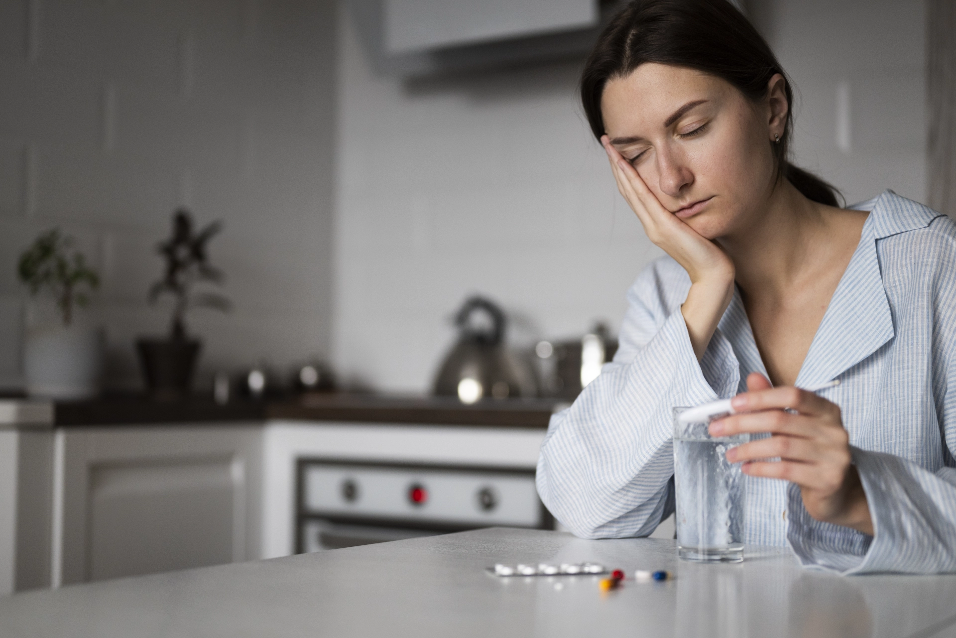
Patients with gonarthritis (inflammation of the knee joint) may also have anemia. This may be a manifestation of the main disease or, in some cases, anemia itself may provoke joint inflammation. For example, in rheumatoid arthritis, blood tests may show anemia, thrombocytosis, and elevated ESR — indicators of disease activity. In such cases, it is not necessary to treat anemia with iron supplements; rather, proper anti-inflammatory therapy to control disease activity and flare-ups is sufficient, and laboratory markers will normalize on their own.
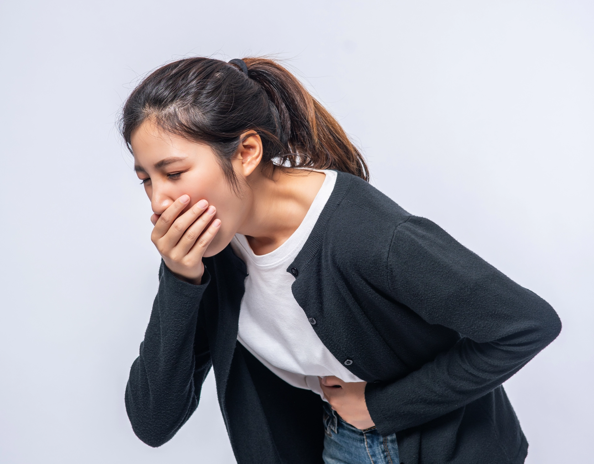
Anemia during inflammatory joint diseases may also be caused by prolonged use of nonsteroidal anti-inflammatory drugs (NSAIDs) such as diclofenac, ibuprofen, aertal, or indomethacin, which can lead to gastritis or peptic ulcers. One possible manifestation of these complications is anemia.
Knee pain can also be occupational. For example, knee injuries are very common among football players, since football involves fast running, sudden stops and turns, jumping, and kicking. Players may stretch or tear cruciate ligaments, damage the menisci, dislocate the kneecap, or develop inflammation or muscle strain.
A deficiency of vitamin D and magnesium can also cause knee and muscle pain. It is particularly important to replenish magnesium deficiency, as all enzymes involved in vitamin D metabolism require magnesium as a cofactor. Even when magnesium levels in bone tissue decrease, its concentration in the blood may remain within the normal range — this is known as chronic latent magnesium deficiency.
What Is Appendicitis?
16 February 2026
Appendicitis is a condition in which the appendix becomes inflamed and swollen. The appendix is a small, finger-shaped pouch attached to the lower right part of the large intestine. Although it is not essential for survival, its inflammation can quickly become dangerous if not treated in time. Appendicitis usually occurs when the appendix becomes blocked by hardened stool, swollen lymphatic tissue or infection. Bacteria multiply inside the blocked appendix, causing swelling, reduced blood flow and tissue damage. If untreated, the appendix may rupture and spread infection into the abdominal cavity. For this reason, appendicitis is considered a medical emergency. It most commonly affects people between 10 and 30 years of age, but it can occur at any age. Signs and Symptoms Early recognition is very important. Typical symptoms include: • Pain that begins near the belly button • Pain that moves to the lower right abdomen • Increasing abdominal pain • Nausea and vomiting • Loss of appetite Other possible symptoms may include: • Fever • Abdominal tenderness • Pain that worsens with movement or coughing • Bloating • Constipation or, in some cases, diarrhea Children, elderly individuals and pregnant women may not have typical symptoms. If the appendix ruptures, severe abdominal pain, high fever and signs of widespread infection may develop. This requires immediate emergency care. Diagnosis A healthcare provider evaluates symptoms, performs a physical examination and may order tests such as: • Blood tests • Abdominal ultrasound • CT scan These help confirm appendicitis and rule out other conditions. Treatment Appendicitis requires urgent treatment. Antibiotics are given to treat and prevent infection. In very early and mild cases, antibiotics alone may temporarily improve symptoms, but recurrence is common. Surgery (appendectomy) is the standard and most reliable treatment. The appendix is usually removed within 24 hours of diagnosis to prevent rupture. Most surgeries are performed laparoscopically, allowing faster recovery. In complicated cases, open surgery may be necessary. The appendix is not a vital organ, and people live normal lives after its removal. Complications and Recovery If untreated, appendicitis may lead to rupture, abscess, peritonitis or sepsis. Early treatment greatly reduces these risks. Most patients recover completely, especially with early care. After uncomplicated surgery, many return home within 24 hours and fully recover within several weeks. Seek immediate medical attention if you experience persistent or worsening pain in the lower right abdomen, especially with fever, nausea or vomiting.
10 Tips to Prevent Breast Cancer
11 February 2026
Breast cancer is influenced by many factors. Some—such as genetics, age, and family history—cannot be changed. However, research shows that healthy lifestyle choices combined with regular screenings can significantly lower risk and improve early detection. Although prevention cannot be guaranteed, proactive steps can reduce your chances of developing breast cancer and increase the likelihood of detecting it early, when treatment is most effective. 1. Maintain a healthy weight Excess weight, especially after menopause, increases risk because fat tissue produces estrogen. Maintain a stable, healthy weight through balanced nutrition and regular activity. 2. Stay physically active Exercise helps regulate hormones and control weight. Aim for 150 minutes of moderate activity or 75 minutes of vigorous exercise weekly, plus strength training twice a week. 3. Limit alcohol Alcohol increases breast cancer risk. If you drink, limit it to one drink per day—or avoid it entirely. 4. Choose a balanced diet There is no single “anti-cancer diet,” but eating fruits, vegetables, whole grains, legumes, and healthy fats supports overall health. Reduce processed meat, red meat, sugary drinks, and heavily processed foods. 5. Avoid smoking Smoking is linked to many cancers, including breast cancer. Quitting greatly benefits overall health. 6. Breastfeed if possible Breastfeeding may slightly reduce risk, especially when continued longer. 7. Use hormones carefully Hormone replacement therapy and some hormonal contraceptives may slightly increase risk. Always discuss options with your doctor and use the lowest effective dose for the shortest time. 8. Know your family history Understand cancer history on both sides of your family. 9. Take extra steps if high-risk High-risk individuals may need earlier screening, preventive medications, genetic counseling, or other preventive strategies. 10. Prioritize regular screening Healthy habits reduce risk—but screening saves lives. Early detection greatly improves outcomes. Screening may include: • Clinical breast exams • Mammography based on age and risk • Breast ultrasound, especially for dense breast tissue • MRI for high-risk individuals Ultrasound is safe, painless, and radiation-free, and can detect changes not always visible on mammograms. If you notice a lump, skin changes, nipple discharge, or breast shape changes, seek medical care promptly. Regular check-ups are not routine—they are protection.
Importance of Vitamin D for Infants
10 February 2026
The first year of life is a time of very rapid growth, and good nutrition is essential for healthy development. Vitamin D is one of the most important nutrients for infants, as it supports strong bones, teeth, and the immune system. Because babies cannot safely get enough vitamin D from sunlight or diet alone, supplementation is often necessary from birth. Vitamin D is sometimes called the “sunshine vitamin” because the body can produce it when skin is exposed to sunlight. Why do babies need vitamin D? Infants grow very quickly during their first 12 months, which increases their need for vitamin D. Without enough vitamin D, calcium cannot be absorbed properly, leading to weak or soft bones. Severe deficiency can cause rickets, a condition that may result in delayed growth, bone pain, and skeletal deformities. Vitamin D also plays an important role in supporting the immune system, helping babies fight infections and stay healthy. Why sunlight and diet are not enough Although sunlight is a natural source of vitamin D, babies have very sensitive skin and should not be exposed to direct sunlight, especially in early life. Breast milk, while the best source of nutrition for infants, usually does not contain enough vitamin D to meet daily needs. Solid foods introduced later are also low in vitamin D. For these reasons, health authorities recommend vitamin D supplementation for many infants. Which babies need vitamin D supplements? • All breastfed babies should receive a daily vitamin D supplement from birth. • Babies who receive both breast milk and small amounts of formula also need supplementation. • Formula-fed babies usually do not need extra vitamin D if they consume enough fortified infant formula. Babies at higher risk of deficiency Babies born to mothers with low vitamin D levels and infants with darker skin tones are more likely to have vitamin D deficiency, as darker skin produces less vitamin D from sunlight. In these cases, regular supplementation is especially important. How to give vitamin D Vitamin D supplements for babies are widely available as liquid drops. They can be given directly into the baby’s mouth using a dropper or oral syringe. Always choose a supplement suitable for infants, follow the instructions carefully, and do not exceed the recommended dose. In summary, vitamin D is essential for healthy growth, strong bones, and immune support in infants. Since babies cannot safely rely on sunlight or diet alone, providing a daily vitamin D supplement during the first year is a simple and effective way to support their long-term health.
Why is gastric ultrasound important before surgery?
06 February 2026
When someone undergoes surgery, one of the biggest hidden risks comes from the stomach. If food or liquid remains in the stomach during anesthesia, it can accidentally travel into the lungs—a dangerous situation known as aspiration. This can lead to serious complications such as breathing difficulties, lung inflammation, pneumonia, or, in severe cases, life-threatening outcomes.To reduce this risk, patients are usually asked to fast before surgery. While fasting rules work well for many people, they don’t always guarantee an empty stomach—especially for those with certain medical conditions. That’s where gastric ultrasound becomes a powerful and reassuring tool.What Is Gastric Ultrasound?Gastric ultrasound is a quick, painless, and non-invasive scan that allows doctors to see what’s inside your stomach in real time. It’s done right at the bedside and takes only a few minutes. Using sound waves, the ultrasound shows whether your stomach is empty or contains food or liquid—and even what kind.This information helps doctors make safer decisions about when and how to perform anesthesia, especially when the situation is unclear.How Gastric Ultrasound Helps Keep You SafeA gastric ultrasound allows healthcare providers to:• Check whether your stomach is empty or full• Identify solid food, thick liquids, or clear fluids• Estimate the amount of liquid in the stomach• Decide whether surgery can proceed safely or should be delayed• Choose the safest anesthesia and airway protection methodFor urgent surgeries, this information is especially valuable—it helps doctors reduce risks even when waiting isn’t an option.Is Gastric Ultrasound Safe?Yes—gastric ultrasound is extremely safe. It does not use radiation and has no known side effects. There are very few situations where it might not be suitable, such as severe abdominal injury or when a patient cannot safely lie on their side.In some cases, special techniques or temporary tubes may be used to empty the stomach and protect the lungs during anesthesia.Gastric ultrasound is a simple exam that can make a big difference in surgical safety. It provides clarity when fasting status is uncertain and helps doctors tailor care to your individual needs. Most importantly, it reduces the risk of serious complications and helps ensure that your procedure is as safe as possible.

Fill in the required fields

I am waiting for a call

We will help you quickly find what you need!

Thank you!
Our employee will contact you soon
Close
dalimed medical
How can we help?
Leave your number and we will call you back
Book a call
I agree with the terms of personal data processing
Thank you!
Our employee will contact you soon
Close