Eyelid Demodicosis — Causes, Symptoms, and Modern Treatment Methods

Eyelid Demodicosis — Causes, Symptoms, and Modern Treatment Methods

Eyelid Demodicosis — Causes, Symptoms, and Modern Treatment Methods
Demodicosis is a common yet often overlooked inflammatory disease of the eyes and eyelids, caused by the excessive multiplication of microscopic mites —

Demodex folliculorum

and

Demodex brevis

— which normally live on the human skin. These mites inhabit the hair follicles and meibomian glands, feeding on skin sebum and dead cells. Under normal conditions they do not cause harm, but when their number exceeds the norm, an inflammatory reaction occurs, leading to irritation, itching, and various ocular surface disturbances.
The activation of these mites is related to both internal and external factors.

The main causes include:

Weakened immune system

due to chronic diseases, infections, or prolonged stress.

Increased skin oiliness

, which creates a favorable environment for mite growth.

Hormonal imbalance

, such as thyroid or ovarian dysfunction.

Age-related changes

, as the condition is more common in middle-aged and elderly people.

Poor eyelid hygiene

and overuse of oily cosmetics, especially mascara and eye creams.
When several of these factors act together, Demodex mites begin to reproduce rapidly, causing inflammation of the eyelids and eye surface.
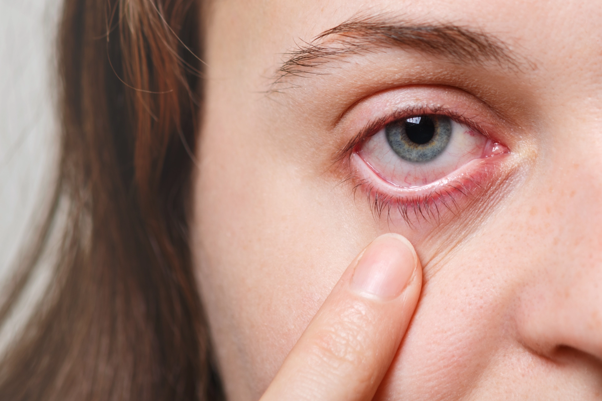
Symptoms

Demodicosis develops gradually. Initially, a person may notice mild itching or tingling around the eyelids, but over time the symptoms become more pronounced.

Typical symptoms include:

• Itching and burning of the eyelids, especially in the morning.
• Redness, swelling, and a feeling of heaviness in the eyelids.
• Yellowish-white crusts or discharge at the base of the eyelashes.
• Tearing, a gritty or foreign body sensation.
• Recurrent inflammation along the eyelid margins, sometimes involving the cornea and conjunctiva.
If the mites affect the meibomian glands, meibomian gland dysfunction may develop, causing chronic eye dryness, irritation, and light sensitivity.

Diagnosis

Diagnosis is confirmed by an ophthalmologist through microscopic examination of eyelashes to identify mites and their eggs. Additionally, the condition of the cornea and tear film is evaluated to determine the severity of inflammation and to detect possible complications.

Modern Treatment Approaches

Treatment of demodicosis is lengthy and requires a complex, combined approach aimed at reducing mite activity, eliminating inflammation, and restoring normal eyelid function.
The main treatment directions are:
1.

Eyelid and eyelash hygiene

— daily cleansing with special lotions or wipes to remove secretions and mite waste; warm compresses to improve meibomian gland function.
2.

Topical therapy

— ointments and drops containing anti-mite components such as tea tree oil, metronidazole, or ivermectin; anti-inflammatory agents to reduce redness and swelling.
3.

Systemic therapy

— oral medications in severe cases, prescribed only under medical supervision.
4.

Supportive therapy

— artificial tears to relieve dryness, as well as lifestyle adjustments including healthy diet, adequate sleep, and stress management.

Prevention

To prevent recurrence, it is essential to maintain regular eyelid hygiene, limit the use of oily cosmetics, and avoid using old or contaminated makeup products. Any early signs such as itching, redness, or tearing should prompt a visit to an ophthalmologist to avoid complications.

Conclusion

Demodicosis does not threaten vision directly, but it can significantly affect quality of life by causing chronic discomfort, irritation, and cosmetic concerns. Early diagnosis, proper hygiene, and comprehensive treatment allow effective control of the disease and help prevent relapses.
What Is Elastography of the Thyroid Gland?
29 January 2026
When you hear the word elastography related to the thyroid, there’s no need to worry. It’s simply a modern imaging method that gives doctors more information than a regular ultrasound. Elastography works like an “electronic palpation” — it allows doctors to assess how soft or stiff the thyroid tissue is, without physically touching it. Why Does This Matter? The thyroid gland is located at the front of the neck and often develops nodules, which are small lumps of tissue. Most thyroid nodules are harmless, but some may need closer attention. Doctors usually evaluate them with standard ultrasound and, in certain cases, a fine-needle biopsy. While these methods are helpful, they don’t always give the full picture. Elastography adds important extra information by showing how stiff the tissue is. Changes in tissue stiffness can occur with conditions such as nodules, inflammation, or other thyroid disorders. This information helps doctors better understand what they are seeing on ultrasound. How Does Thyroid Elastography Work? Elastography is performed during an ultrasound examination and does not cause pain or discomfort. The technique measures how thyroid tissue responds to gentle pressure or sound waves. In simple terms, elastography helps to: • Compare softer and stiffer areas within the thyroid • Provide additional details alongside regular ultrasound images • Support doctors in evaluating thyroid nodules more accurately What Are the Benefits for Patients? • Non-invasive and painless • Performed together with standard thyroid ultrasound • Provides additional information without extra procedures Where Can You Do This Exam? If you may benefit from thyroid elastography — especially if a previous ultrasound showed nodules or changes in the thyroid — you can have this examination at Dalimed Medical Center. The medical team performs thyroid elastography as part of comprehensive ultrasound diagnostics and explains the results in a clear and patient-friendly way.
What is a Holter monitor?
26 January 2026
A Holter monitor is a small, wearable medical device used to continuously record the electrical activity of your heart over an extended period, usually 24 to 48 hours, and sometimes longer. It’s a type of ambulatory electrocardiogram (ECG/EKG), meaning it monitors your heart while you go about your normal daily routine outside of a clinic or hospital. Unlike a standard ECG, which captures your heart’s activity for just a few minutes while you’re resting, a Holter monitor provides a much broader picture. By recording every heartbeat over one or more days, it helps healthcare providers detect heart rhythm problems that may appear only occasionally or during specific activities such as exercise, stress or sleep. How a Holter monitor works Your heart beats because of natural electrical impulses that control the timing and rhythm of contractions. A Holter monitor records these impulses through electrodes placed on your chest. The system usually includes: • Electrodes – small adhesive patches attached to the skin that detect electrical signals • Wires – connect the electrodes to the recording device (some newer models are wireless patch-style units) • Recording device – a lightweight, battery-powered monitor worn on a belt, strap or in a pocket The device stores all the data while you wear it. Your healthcare provider reviews the recordings only after you return the monitor—there’s no real-time monitoring. Why a Holter monitor may be needed Your healthcare provider might recommend a Holter monitor if symptoms suggest a heart rhythm issue that isn’t visible on a resting ECG. It’s especially useful when symptoms are intermittent. Common reasons include: • Dizziness, fainting or unexplained fatigue • Heart palpitations or irregular heartbeats • Chest discomfort or shortness of breath • Evaluating how well heart medications or a pacemaker are working • Assessing heart rhythm changes after a heart attack or in certain heart conditions Because the monitor records continuously, it increases the chance of capturing abnormal rhythms such as very fast, very slow or irregular heartbeats. What to expect while wearing a Holter monitor Getting set up is simple and usually done on an outpatient basis. After the electrodes or patch are placed on your chest, you can return to most of your normal activities, including work and light exercise, unless your provider advises otherwise. You’ll likely be asked to: • Keep a diary of activities and symptoms (noting time, activity and how you felt) • Avoid getting the device wet (no showering, bathing or swimming) • Stay away from strong magnetic or high-voltage environments that could interfere with the recording Recording what you’re doing when symptoms occur helps your provider match those moments with changes seen in your heart rhythm. Risks and limitations A Holter monitor is safe and painless. Risks are minimal, but some people may notice: • Mild skin irritation or itching where electrodes are attached • Discomfort when electrodes are removed • Loosening of electrodes due to sweating Electrical appliances, magnets or cell phones placed too close to the device may interfere with signal quality, so following instructions is important. After the monitoring period Once the monitoring time is complete, you return the device to your healthcare provider. The data from the monitor, along with your activity diary, is analyzed to look for abnormal heart rhythms, pauses or unusual rate changes. Results may show normal heart activity or detect rhythm disorders such as atrial fibrillation, bradycardia (slow heart rate), tachycardia (fast heart rate) or extra beats. Your provider will explain the findings and discuss next steps, which may include additional tests, medication adjustments or other treatments if needed. A Holter monitor is a valuable, noninvasive tool that helps healthcare providers better understand how your heart behaves throughout your everyday life—not just in a brief moment in the exam room. If you have heart-related symptoms, you can visit Dalimed Medical Center for a specialist consultation and, if needed, undergo Holter monitoring using modern equipment in a comfortable clinical setting.
What is human cytomegalovirus?
19 January 2026
Human cytomegalovirus (HCMV), commonly known as CMV, is a widespread virus from the beta-herpesvirus family that causes lifelong infection. Once a person is infected, the virus remains in the body in a dormant state and may reactivate later. CMV is extremely common worldwide, with infection rates ranging from 55% to nearly 100%, depending on age, geography, and socioeconomic conditions. Most healthy people experience no symptoms or only mild, flu-like illness during primary infection. However, CMV can cause severe and life-threatening disease in individuals with weakened immune systems, including newborns, pregnant women, organ transplant recipients, and people living with HIV/AIDS. CMV and High-Risk Groups CMV is the leading infectious cause of congenital abnormalities in developed countries, affecting approximately 1–2.5% of all live births. The virus can cross the placenta during pregnancy and infect the fetus, even if the mother has no symptoms. Complications of congenital CMV may include: • Low birth weight • Hearing and vision loss • Microcephaly • Enlarged liver and spleen • Developmental and cognitive delays In immunocompromised adults, CMV can cause pneumonia, gastrointestinal disease, retinitis, neurological complications, and may contribute to transplant rejection. Transmission of CMV CMV spreads through direct contact with infected body fluids, such as saliva, urine, blood, breast milk, and sexual fluids. Young children are a common source of infection due to prolonged viral shedding. Main routes of transmission include: • Close contact with saliva or urine • Sexual contact • Breastfeeding • Blood transfusions and organ transplants Diagnosis, Treatment, and Prevention CMV is diagnosed using blood, urine, or saliva tests. Treatment with antiviral medications (ganciclovir or valganciclovir) is usually limited to high-risk patients, as these drugs cannot reverse existing damage and may cause side effects. Prevention focuses on reducing exposure, especially during pregnancy:• Strict hand hygiene • Avoiding sharing utensils with young children • Practicing safe sex • Medical monitoring after transplantation Conclusion Human cytomegalovirus is usually harmless in healthy individuals but can cause serious disease in vulnerable populations. Improved understanding of CMV biology and immune interactions is essential for better therapies and the development of an effective vaccine.
What is malnutrition?
16 January 2026
Malnutrition: what it means and why it matters Malnutrition is a condition that occurs when the body does not receive the correct amount or balance of nutrients needed to stay healthy. This problem is not limited to hunger alone. A person may eat too little, eat too much, or consume foods that lack essential vitamins and minerals. Any long-term mismatch between nutritional needs and intake can harm the body’s normal functions. Malnutrition is broadly divided into undernutrition and overnutrition. Undernutrition develops when the body lacks sufficient energy, protein or micronutrients to maintain tissues, immunity and growth. It may cause weight loss, muscle wasting, fatigue, frequent infections and delayed physical or intellectual development in children. Overnutrition occurs when excessive calories or nutrients are consumed over time, often leading to obesity and metabolic disorders such as diabetes, heart disease and high blood pressure. Types of malnutrition • Undernutrition – deficiency of calories, protein, vitamins or minerals due to poor intake, impaired absorption or increased nutritional needs. • Overnutrition – excessive intake of calories or nutrients, including unhealthy weight gain and, in rare cases, toxicity from supplements. Malnutrition can affect people of all ages, but children, older adults, individuals with chronic illnesses and those with limited access to nutritious foods are particularly vulnerable. Importantly, malnutrition is not always visible — a person may appear overweight while still lacking essential micronutrients. Main causes of malnutrition • Unbalanced or insufficient diet, often linked to poverty, food insecurity or lack of nutritional knowledge. • Medical and lifestyle factors, such as chronic disease, digestive disorders, sedentary habits, mental health conditions or improper use of supplements. Diagnosis is based on dietary assessment, physical examination and, when needed, laboratory tests to identify nutrient deficiencies or excesses. Treatment depends on the type and severity of malnutrition and may involve dietary changes, nutritional supplementation, medical monitoring or long-term lifestyle support. Preventing malnutrition starts with a varied, balanced diet that meets the body’s nutritional needs. Early recognition and appropriate intervention play a key role in reducing long-term health complications and improving overall quality of life.

Fill in the required fields

I am waiting for a call

We will help you quickly find what you need!

Thank you!
Our employee will contact you soon
Close
dalimed medical
How can we help?
Leave your number and we will call you back
Book a call
I agree with the terms of personal data processing
Thank you!
Our employee will contact you soon
Close